musculoskeletal radiology in Surrey
Specialist Musculoskeletal MRI and CT Imaging for Sports Injuries, Joint Pain and Orthopaedic Conditions
We provide specialist imaging of bones, joints, muscles, tendons and ligaments. Our expert reporting supports the diagnosis of sports injuries, arthritis, persistent pain and complex orthopaedic conditions, helping patients and clinicians make informed decisions about the next stage of care.
What Is Musculoskeletal Imaging?
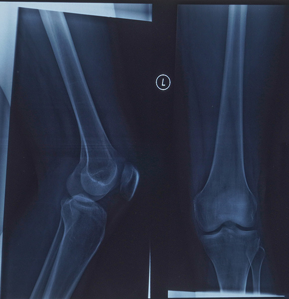
Musculoskeletal imaging uses a variety of imaging methods to examine the structures that enable movement and support the body. These can be assessed with x-rays (including CT), magnetic resonance imaging (MRI) or ultrasound.
The different types of imaging give diAerent information so sometimes different scan types are combined to give detailed information about bone, cartilage, joints, muscles, tendons, ligaments and surrounding soft tissues. We use the most suitable examination for the clinical question, then provide a detailed consultant radiologist report to support diagnosis, treatment planning and rehabilitation.
What Can Musculoskeletal Imaging Detect?
High-quality joint imaging and orthopaedic imaging can help identify a wide range of injuries and conditions, including subtle abnormalities that may not be clear on examination alone. Depending on the area being assessed, imaging may help detect:
Tendon and Ligament Injuries
Inflammation, degeneration, sprains, partial tears and complete ruptures affecting tendons and ligaments.
Cartilage and Joint Damage
Cartilage wear, focal defects, meniscal injuries, joint instability and other abnormalities within weight-bearing joints.
Bone Injuries and Abnormalities
Fractures, stress fractures, bone bruising, structural abnormalities and complex bony injuries.
Arthritis and
Degenerative Change
Osteoarthritis, inflammatory arthritis, joint degeneration and changes associated with pain, stiffness and reduced movement.
Muscle and
Soft-Tissue Conditions
Muscle strains, tears, haematomas, chronic muscle problems and soft-tissue masses requiring further assessment.
Spinal Conditions
Disc disease, nerve compression, facet joint changes and degenerative conditions affecting the neck or back
Common Applications of Musculoskeletal Imaging

Sports Injury Imaging
MRI is particularly valuable for assessing sporting injuries involving the knee, shoulder, hip, ankle and muscles. It can help identify ACL injuries, meniscal tears, muscle strains, rotator cuff tears and tendon damage, supporting an appropriate return-to-sport or rehabilitation plan.

Persistent Joint Pain
Joint pain may arise from cartilage wear, inflammation, tendon disease, joint instability or an injury that has not healed as expected. Specialist imaging can help identify the source of ongoing symptoms and guide the most appropriate treatment pathway.

Arthritis Imaging
Imaging plays an important role in assessing arthritis and monitoring its progression. We can identify changes associated with osteoarthritis, inflammatory arthritis and joint degeneration, helping clinicians distinguish between different causes of pain, stiffness and reduced movement.

Spine Assessment
Musculoskeletal MRI can provide detailed assessment of the spine, including discs, vertebrae, facet joints, nerves and surrounding soft tissues. It is commonly used for back pain, neck pain, suspected nerve irritation, sciatica and degenerative spinal conditions.

Complex Fractures and Surgical Planning
CT imaging offers excellent bone detail and can be particularly useful when evaluating complex fractures, joint surfaces and bony alignment. It may also support surgical planning by helping orthopaedic teams understand the exact nature and extent of an injury.
Why Choose Musculoskeletal Imaging?
Accurate imaging can reduce uncertainty and help avoid unnecessary delays in treatment. By showing the condition of soft tissues, joints and bones in greater detail, MRI and CT can assist clinicians in deciding whether physiotherapy, medication, injections, further specialist review or surgery may be appropriate. Imaging findings are always considered alongside symptoms, examination and medical history.
Need specialist imaging for a sports injury, persistent joint pain or an orthopaedic condition?
Refer to SHB Medical Imaging for expert MRI or CT assessment and detailed consultant reporting.
How to Prepare for Your Musculoskeletal Imaging Scan
Preparation depends on the type of examination being performed. Most musculoskeletal MRI scans require no special preparation, although you must tell us about any implants, metal devices, previous surgery or possibility of pregnancy.
For CT examinations, we will advise if contrast is required and whether any specific instructions apply. Please wear comfortable clothing and bring relevant previous imaging or clinical information where requested.
Working Across Leading Hospitals
We provide musculoskeletal radiology in Surrey, Hampshire and Berkshire patients can access through leading local hospital facilities. Our specialist reporting supports local clinicians, helping patients receive expert assessment close to home. Scan availability may vary by site and examination type.
Frimley Park Hospital
Portsmouth Road,
Frimley,
Camberley,
GU16 7UJ
Aldershot Centre for Health
Hospital Hill,
Aldershot,
GU11 1AY
Clare Park Hospital
Crondall Lane,
Farnham,
GU10 5XX
Heatherwood Hospital
Brook Avenue,
Ascot
SL5 7GB
Wexham Park Hospital
Wexham Street,
Slough,
SL2 4HL
Questions about musculoskeletal radiology in Surrey
What is a Cardiac CT scan?
A cardiac CT scan is a specialised X-ray examination that produces detailed images of the heart and coronary arteries. Also called coronary CT angiography or CTCA, it may use an iodine-based contrast injection to show the blood vessels supplying the heart.
What can a Cardiac CT scan diagnose?
Cardiac CT can help assess coronary artery narrowing, calcified and non-calcified plaque, coronary artery disease, congenital coronary variations and cardiac anatomy. It may also identify relevant incidental findings in visible areas of the chest.
Who should consider having a private Cardiac CT scan?
A private cardiac CT scan may be suitable for people with chest pain, breathlessness, cardiovascular risk factors, inconclusive cardiac test results or a need for coronary calcium scoring. Suitability should always be reviewed by a GP, cardiologist or other appropriate clinician.
Can a Cardiac CT scan detect blocked or narrowed coronary arteries?
Yes. Coronary CT angiography can provide detailed images of the coronary arteries and help identify plaque or narrowing that may reduce blood flow to the heart muscle. Your clinician will use the results to decide whether further assessment or treatment is needed.
Do I need a GP or consultant referral for a private Cardiac CT scan?
A referral from a GP, consultant or appropriate healthcare professional is usually required so the examination can be clinically reviewed and tailored to your symptoms. SHB Medical Imaging may discuss self-referral options in some circumstances, subject to clinical suitability.
How should I prepare for a Cardiac CT scan?
Preparation depends on the type of cardiac CT examination. You may be asked to avoid caffeine, smoking and strenuous exercise beforehand. Tell the team about allergies, asthma, kidney problems, diabetes, pregnancy and any previous contrast reaction, and follow all medication instructions provided.
How long does a Cardiac CT scan take?
The image acquisition itself is usually very quick and may be completed within a single heartbeat using advanced cardiac CT protocols. Your overall appointment will be longer to allow for registration, preparation, possible heart-rate medication and contrast administration where required.
Is a Cardiac CT scan safe and does it involve radiation?
Cardiac CT uses X-rays and therefore involves radiation exposure. SHB Medical Imaging uses optimised low-dose cardiac CT protocols to keep the dose as low as reasonably achievable while maintaining diagnostic image quality. Contrast use is assessed carefully for each patient.
How quickly will I receive the results of my Cardiac CT scan?
Your images are reviewed by a consultant radiologist with relevant cardiac imaging expertise, and a detailed report is sent to your referring clinician. They will discuss the findings and any next steps with you. Contact SHB Medical Imaging for current reporting times.
What is the difference between a Cardiac CT scan and a Cardiac MRI scan?
Cardiac CT uses X-rays to provide particularly detailed images of the coronary arteries, calcium and cardiac anatomy, often very quickly. Cardiac MRI uses magnetic fields rather than radiation and is commonly used to assess heart muscle function, tissue characteristics and some structural conditions.
Why choose a private Cardiac CT scan instead of waiting for an NHS appointment?
Private cardiac CT can offer access to local hospital-based imaging in Surrey, Hampshire and Berkshire, with consultant reporting and advanced scanner technology. Availability varies, but private access may help patients obtain an appropriate clinically reviewed scan without unnecessary travel.
Refer for Specialist Musculoskeletal Imaging
Whether you are investigating a recent injury, chronic joint pain, arthritis symptoms or a complex orthopaedic condition, we can help provide the detailed imaging and specialist interpretation needed for confident next steps. SHB Medical Imaging supports referrals from GPs, consultants, physiotherapists, sports medicine clinicians and other healthcare professionals.
To discuss a referral or our diagnostic imaging services in Surrey, please contact us today.
TBC