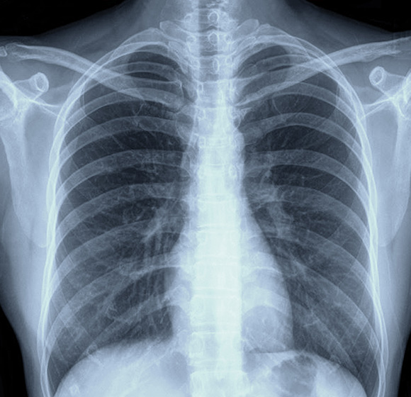
Thoracic Imaging in Surrey
Consultant-Led Private Thoracic Imaging and Chest CT
We provide specialist thoracic imaging for patients across Surrey, Hampshire and Berkshire. Our consultant-led service uses advanced imaging technology to assess the lungs, airways, chest wall and surrounding structures, helping clinicians investigate symptoms, monitor known conditions and plan appropriate care.
What Is Thoracic Imaging?
Thoracic imaging is the assessment of structures within the chest, including the lungs, pleura, airways, chest wall, mediastinum, heart and major blood vessels. Depending on the clinical question, this may involve a chest CT scan or another relevant examination. Our thoracic radiology service combines carefully selected imaging protocols with interpretation by experienced consultant radiologists, providing detailed, clinically useful information for referrers and patients.
What Can a Cardiac CT Scan Detect?
A cardiac CT scan can provide important information about the coronary arteries, heart anatomy and surrounding structures. It is commonly used to investigate whether narrowing or disease within the coronary arteries may be contributing to symptoms or cardiovascular risk.
Depending on the type of examination requested, cardiac CT may help identify:
Lung Nodules and Masses
Small lung nodules, larger masses or other areas that may require further assessment, comparison or follow-up imaging.
Infection, Inflammation
and Scarring
Changes associated with chest infections, inflammation, fibrosis, scarring and other diffuse lung abnormalities.
Airway and Lung Conditions
Airway narrowing, bronchiectasis, emphysema and changes associated with interstitial lung disease or other respiratory conditions.
Pulmonary Embolism and Blood Vessel Concerns
Where clinically appropriate, contrast-enhanced CT pulmonary angiography can assess for blood clots in the arteries of the lungs.
Pleural, Mediastinal and Lymph Node Abnormalities
Fluid around the lungs, pleural thickening, enlarged lymph nodes, mediastinal masses and other changes in the central chest.
Thoracic Cancer Assessment and Monitoring
Findings associated with known or suspected cancer affecting the lungs, pleura or mediastinum, including changes in response to treatment.
Imaging findings are interpreted in the context of the referral information and should be discussed with the referring clinician.
Common Applications of Thoracic Imaging

Lung Nodules
Lung nodules are small areas seen within the lung tissue. Thoracic imaging can characterise their size, location and appearance, and provide a baseline for follow-up where recommended by the referring clinician or relevant clinical guidance.

Interstitial Lung Disease
High-quality lung imaging can help assess patterns of inflammation, fibrosis or scarring in the lungs. This supports the investigation and monitoring of interstitial lung disease, alongside clinical review, lung function testing and other assessments.

Pulmonary Embolism Assessment
A CT pulmonary angiogram may be requested when there is a clinical concern about a blood clot in the arteries of the lungs. This examination uses contrast material and is only arranged when appropriate following clinical assessment.

Thoracic Oncology Imaging
Chest CT can assist in the assessment of suspected or known cancer affecting the lungs, pleura or mediastinum. It can help clinicians evaluate the extent of disease, assess treatment response and identify changes over time.

Mediastinal Pathology
The mediastinum is the central area of the chest containing major blood vessels, lymph nodes, nerves and other structures. Mediastinal imaging can investigate enlargement, masses, lymph node changes or other abnormalities identified through symptoms or previous tests.
Why Choose Thoracic Imaging?
Thoracic imaging provides detailed views of the chest in a relatively short examination. A chest CT can demonstrate the lungs, airways and surrounding tissues with far greater anatomical detail than many other imaging methods. This can support earlier diagnosis, guide onward referral and help clinicians monitor known conditions. The precise examination requested will depend on your symptoms, medical history and the question your referring clinician needs answered.
Need specialist thoracic imaging or a chest CT scan?
Contact us to discuss your referral, the most suitable examination and local appointment availability.
Why Choose SHB Medical Imaging?
Advanced CT Technology
We have access to latest-generation CT scanner platforms from Canon at Frimley Park Hospital and GE Healthcare at Heatherwood and Wexham Park Hospitals, supporting detailed thoracic radiology examinations.
Optimised Imaging Protocols
Our protocols are developed by consultant radiologists for the clinical question being investigated. We tailor examinations rather than relying solely on generic manufacturer settings.
High-Quality Images
Modern scanner technology helps deliver clear images of the lungs, chest wall and mediastinum. This supports confident reporting and appropriate clinical decision-making.
Dose-Conscious Imaging
We aim to use the lowest radiation dose reasonably achievable while maintaining the image quality required for accurate chest diagnostics.
Considered Contrast Use
Where contrast is required, protocols are designed to use an appropriate amount for the examination. This may be particularly relevant for patients where contrast use requires additional consideration.
Consultant-Led Reporting
Your images are reviewed by a consultant radiologist with relevant expertise in chest imaging, cardiac imaging in Surrey or the particular clinical area being assessed.
NHS Expertise, Private Access
Our radiologists combine NHS consultant experience with accessible private imaging pathways, helping local patients access specialist assessment without unnecessary travel.
Local Hospital-Based Care
SHB Medical Imaging provides cardiothoracic imaging in Surrey and the surrounding Hampshire and Berkshire areas through established hospital-based locations.
The Patient Journey for
Thoracic Imaging

Referral Received
We receive a referral from your consultant, GP or another appropriate healthcare professional. In certain circumstances, we may also be able to advise patients about self-referral options.


Clinical Review and Appointment
We review the referral to confirm that the requested examination is appropriate. Our team will then arrange an appointment at the most suitable available location.

Preparation and Imaging
Before your appointment, we explain what to expect and provide any relevant preparation instructions.
Your scan is performed using a specialist protocol tailored to the clinical question

Consultant Interpretation
A consultant radiologist reviews your images alongside the clinical details provided by your referrer. This ensures the report addresses the reason for the examination.

Comprehensive Report Delivered
A detailed report is issued to the referring clinician, supporting timely decisions about diagnosis, treatment, monitoring or onward referral where required.
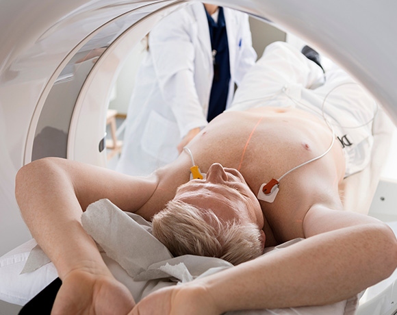
How to Prepare for Your Thoracic Imaging Scan
Preparation depends on the type of thoracic imaging requested. Some chest CT examinations require no special preparation, while others may involve contrast material and require specific instructions. You should tell us and your referrer about allergies, kidney problems, diabetes, pregnancy or the possibility of pregnancy, and any medicines you take. Please bring relevant previous imaging information if requested. Our team will confirm your individual instructions before the appointment.
Working Across Leading Hospitals
SHB Medical Imaging offers specialist lung imaging, chest CT and thoracic radiology services across Surrey, Hampshire and Berkshire. The most appropriate site will depend on the requested examination, your clinical needs and scanner availability.
Frimley Park Hospital
Portsmouth Road,
Frimley,
Camberley,
GU16 7UJ
Aldershot Centre for Health
Hospital Hill,
Aldershot,
GU11 1AY
Clare Park Hospital
Crondall Lane,
Farnham,
GU10 5XX
Heatherwood Hospital
Brook Avenue,
Ascot
SL5 7GB
Wexham Park Hospital
Wexham Street,
Slough,
SL2 4HL
Questions about thoracic imaging in Surrey
What is thoracic imaging?
Thoracic imaging assesses structures within the chest, including the lungs, airways, pleura, chest wall, mediastinum, heart and major blood vessels. It may involve a chest CT scan or another appropriate examination, depending on the clinical question.
When should I consider private thoracic imaging?
Private thoracic imaging may be considered when you have symptoms, an abnormal chest X-ray, a known lung condition requiring monitoring or a clinical concern that needs specialist chest imaging. Your GP, consultant or other healthcare professional can advise whether imaging is appropriate.
What symptoms may require specialist chest imaging?
Specialist chest imaging may be requested for persistent cough, breathlessness, chest discomfort, suspected infection, unexplained symptoms, or abnormal findings from a chest X-ray or previous scan. Imaging is arranged based on clinical assessment rather than symptoms alone.
What can thoracic imaging help diagnose?
Thoracic imaging can help clinicians investigate lung, airway, pleural, mediastinal and chest-wall abnormalities. It can provide detailed information to support diagnosis, treatment planning, follow-up and onward referral, alongside your medical history and other tests.
What lung and chest conditions can thoracic imaging detect?
A chest CT or other thoracic examination may identify lung nodules, infection, inflammation, fibrosis, interstitial lung disease, bronchiectasis, pleural fluid, enlarged lymph nodes, mediastinal abnormalities and changes associated with known or suspected cancer.
Do I need a GP or consultant referral for private thoracic imaging?
A referral from a GP, consultant or another appropriate healthcare professional is usually required for private thoracic imaging. SHB Medical Imaging may also be able to advise on self-referral options in certain circumstances, subject to clinical review.
How quickly can I book a private thoracic imaging appointment?
Appointment times depend on the required scan, clinical review, location and scanner availability. Once a referral has been received and assessed, the team can discuss the earliest suitable appointment at a hospital location in Surrey, Hampshire or Berkshire.
Will my scan be reviewed by a specialist thoracic radiologist?
Your imaging is reviewed by a consultant radiologist with relevant expertise in chest imaging, cardiac imaging or the clinical area being assessed. A detailed report is then sent to your referring clinician.
What should I expect during a thoracic imaging examination?
The examination is performed using a protocol tailored to the reason for your scan. Some chest CT scans require no special preparation, while contrast-enhanced examinations may involve an injection and additional instructions. The team will explain what to expect before your appointment.
Can thoracic imaging help assess persistent cough, chest pain or shortness of breath?
Yes. Where clinically appropriate, thoracic imaging can help investigate persistent cough, breathlessness or chest discomfort by providing detailed views of the lungs and surrounding chest structures. Your referrer will determine whether chest CT or another examination is suitable.
Why choose private thoracic imaging instead of waiting for an NHS appointment?
Private thoracic imaging may offer a local, consultant-led route for patients and referrers seeking an appointment outside standard NHS pathways. SHB Medical Imaging provides tailored protocols, hospital-based scanning and specialist reporting across Surrey, Hampshire and Berkshire.
Make a Referral for Thoracic Imaging
If you are a clinician seeking a thoracic imaging specialist in Surrey, or a patient looking for private chest imaging in Berkshire or Hampshire, please contact SHB Medical Imaging to discuss the referral process. We can help confirm the most appropriate examination, location and appointment arrangements.
Referrals should include relevant clinical history and the reason for the scan, enabling our consultant radiologists to provide a focused and useful report.
TBC